Targeting protein translation and developing ribosome-directed therapy for defeating malaria parasites
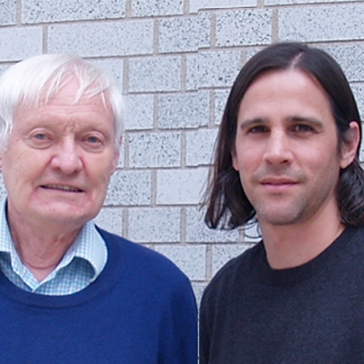
Malaria is an important global pathogen with 100-200 million infections per year, causing almost one million deaths – mostly in the pediatric population and pregnant women. The rates of resistance to all known antimalarials are rising across the globe, and there is an urgent need to develop new drugs for malaria. Drs. Joachim Frank, Professor of Biochemistry and Molecular Biophysics Columbia University, and Jeffrey Dvorin, Assistant Professor of Pediatrics at Harvard Medical School, combine their expertise to conduct a comprehensive study of protein synthesis in Plasmodium falciparum, the parasite responsible for the most severe forms of malaria in humans. Targeting protein translation and the ribosome in this parasite, the two collaborators hope that their research will lead to a precise design of more effective drugs against the disease.
There are currently very few groups looking at the protein synthesis machinery in these parasites or in other tropical parasites, and funding in this area of research is limited. Despite this, Drs. Frank and Dvorin have joined efforts to deliver solutions to global regions where highest technological advancements are most critical but there is less dedicated infrastructure to research. While Dr. Dvorin is an expert on the molecular and genetic pathogenesis of the malaria parasite, Dr. Frank is an expert on structural biology, specializing in 3D reconstruction and cryogenic electron microscopy, or cryo-EM, to probe the structure and function of ribosomes. Recent advances in instrumentation and computer software have made it possible to obtain near-atomic resolution of samples purified from the parasite with this technique, and together, Drs. Frank and Dvorin combine the enormous power of structural knowledge with the ability to directly test molecular hypotheses in live malaria parasites.
Current research topics include:
- What are the structural differences between parasite ribosomes and the host, human ribosomes -- and how can they be exploited for targeting? Like all living organisms, the parasite uses ribosomes, or complex molecular machines found in thousands of genetic blueprints in each cell, to make copies of the proteins it requires to sustain life processes in the cell. Small molecules termed antibiotics can be made to interfere with this process, as they lodge onto the ribosome, effectively killing the parasite. However, an important hurdle is that many of these drugs will interfere with protein synthesis in humans as well. The principle by which proteins are made is the same in all life forms, and targeting the parasite without harming the human host requires very detailed structural and genetic knowledge of the ribosomes in both Plasmodium falciparum and Homo Sapiens – their atomic structure and the precise way they function in synthesizing proteins. Drs. Frank and Dvorin are therefore studying the structure of ribosomes to develop drugs that effectively kill only the parasite, leaving the human host unharmed.
- Are there structural differences in the protein translation machinery and the blood stages in mosquito transmission? The malaria parasite goes through several life stages where it behaves completely differently from one stage to another. Part of these stage differences manifest in the way that protein synthesis is done, and there are currently very few malaria medicines that target parasites at these transmission stages. By understanding the blood stages, Drs. Frank and Dvorin hope to kill both the parasite in an infected individual’s blood as well as the parasites that are transmitted to mosquitos to infect other people.
- Synergy between the Labs: Drs. Frank’s and Dvorin’s groups both have very specialized skill sets: Dr. Dvorin’s group in molecular and genetic manipulation of live parasites, and Dr. Frank’s group in 3D imaging of biological molecules. Ribosomes within the malaria parasite grown and genetically modified in one of its of life cycles and alternate hosts by the Dvorin Lab, can be further examined under high resolution electron microscopy provided by the Frank Lab to understand structure and dynamics of protein synthesis in the parasite. Dr. Frank’s lab is one of the best in the world for structural analysis using cryo-electron microscopy, and the two labs are able to seek feedback from each other, creating a unique synergy to design and redesign experiments.

Bio
Dr. Joachim Frank has been fascinated by the natural world ever since he was a little boy. He conducted experiments in the cellar, then started to take apart and rebuild radios. Physics was therefore a natural choice for him as an undergraduate, and this led him to explore electron microscopy. He found a mentor -- an X-ray crystallographer -- who was convinced that electron microscopy could be used to image biological molecules in 3D. After receiving his Ph.D. on methods of image processing, Dr. Frank developed a large software package powerful enough to reconstruct a molecule from thousands of very noisy images. The ribosome, because of its stability, large size, and strong contrast, was an ideal test object. At a crucial point when he could demonstrate that these reconstructions were better than any previous visualizations, he decided to study the ribosome and the structural basis of protein synthesis full throttle. In this way, Dr. Joachim made many contributions to the understanding of the way the ribosome works in bacteria and eukaryotes. Most recently, as the technique became capable of reaching resolutions that allow atomic modeling, he became interested in looking at the ribosomes of parasites such as Plasmodium falciparum, with the hope to be able to make a contribution toward effective malaria drug design.
http://franklab.cpmc.columbia.edu/franklab
Dr. Jeffrey Dvorin has been interested in how things work for as long as he can remember. He initially studied physics, but later focused his attentions on biological systems. He thought that biological systems – both human and the parasites that invade them – were the most interesting systems to study and to learn how they work. After completing medical school and a Ph.D. in molecular biology, he trained as a Pediatrician and later as an Infectious Diseases specialist. In his research, Dr. Dvorin has relied on genetics and molecular biology to understand important global pathogens. In his Ph.D., he studied HIV and later applied his molecular tool kit to the malaria parasite. http://www.childrenshospital.org/researchers/jeffrey-dvorin
In the News
NIH commits to 78 awards to support exceptional innovation in biomedical research.


