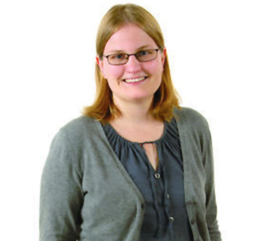
New methods to 'see' biology in action are changing how we think about human health and disease
Optical imaging tools have revolutionized our understanding of living systems by enabling researchers to "peer" into tissues and cells and visualize biological features in real time. While powerful, these probes have been largely confined to monitoring cellular behaviors on a microscopic level. Visualizing cellular interactions and functions across larger spatial scales--including those involved in cell migration to distant tissues, immune function, and other biological processes--remains a daunting task. Dr. Jennifer Prescher, of the University of California, Irvine is developing general toolsets to image such macroscopic cellular networks and behaviors. Her work allows for the development of new chemical tools and noninvasive imaging strategies that address the void of being able to see biological features in real time and therefore inspires new discoveries in a broad spectrum of fields.
Dr. Prescher's young lab, having only been in development for four years, has already achieved important milestones related to imaging probe development, including constructing new tools and performing preclinical analyses. Dr. Prescher and her group are currently translating many of the image probe tools into models of infection and cancer progression. While Dr. Prescher's background is rooted in the molecular underpinnings of big biological systems, it takes a team that is blended in order to design and implement effective imaging tools. Her multidisciplinary team, which bridges the biological, chemical, and physical sciences in addition to engineering, is composed of graduate students, postdocs, and undergraduates. Her collaborations across the country and with other scientists at the University of California, Irvine, are improving the imaging probes and strategies used for routine biomedical analyses. Therefore, Dr. Prescher's research feeds many discoveries by applying the old adage, "seeing is believing." It is through her research that scientists are able to truly spy on cells and tissues in order to improve health, treat disease, and sustain life.
Current research efforts are focused in three areas:
-
Multicellular Tracking: Dr. Prescher is generating novel bioluminescent imaging probes for multi-cellular tracking. She hopes to have a whole suite of different probes for looking at cells and other biological features in live tissues and organisms. Because scientists have run out of tools to easily distinguish cells and other features in living systems, this has thus far been a challenge. Dr. Prescher's innovative approach builds on the incredible examples found in nature to create synthetic versions.
-
Visualizing Cell-to-Cell Contacts In Vivo: Many critical aspects of human biology are driven by communication between cells. Trying to understand these intricate networks without perturbing the environment they are in has been difficult. Dr. Prescher is tackling this challenge by noninvasively imaging cell-to-cell interactions without the need for biopsied tissue or surgical intervention. This work has important ramifications for visualizing metastatic tumors, immune cell function, and neurological disorders, and provides a new vantage point for seeing unique facets of human biology.
-
Biocompatible Chemistries: Dr. Prescher is developing biocompatible chemistries for multi-component imaging. Many biomolecules, like lipids, neurotransmitters, and carbohydrates, cannot be easily studied using classical genetic or molecular biology approaches. Dr. Prescher is developing chemical methods that can more efficiently target the entities with visual probes. These approaches rank among the best approaches for imaging and studying key biomolecules relevant to human health and disease.
Bio
Dr. Prescher earned her Bachelor of Science degree in Chemistry and Mathematics from the University of Wisconsin-La Crosse. She later received her Ph.D. in Chemistry from the University of California, Berkeley, working with Professor Carolyn Bertozzi. Following her doctoral studies, Jenn conducted postdoctoral research with Professor Christopher Contag at Stanford University. She joined the faculty at UC Irvine in 2010.
Dr. Prescher grew up in rural Minnesota where she was always curious about the world around her. Why is the sky blue? Where does the wind come from? What makes the crops grow? As she started to find answers to these questions in school, her excellent teachers exposed her to science - a whole field dedicated to exploring the world, uncovering knowledge, and creating new discoveries. Dr. Prescher loved to learn, and science therefore provided a way to answer the "whys" and "hows" about the world. Her interest in the chemical and molecular sciences, in particular, began in high school and grew exponentially in college. Chemistry and chemical thinking explained a lot about the world around her. After her first exposure to research as an undergraduate, she was convinced that research would be her path. As an undergraduate, Dr. Prescher synthesized hallucinogen analogs that were used to map the structures of serotonin receptors in the brain. Today, her research is still motivated by a love for learning and trying to use chemical methods and molecular thinking to unravel biological mysteries.
In her free time, aside from research Dr. Prescher is a sports enthusiast. She loves to play and watch sports and especially enjoys cheering on her teams from Minnesota, the Vikings and the Twins.
Website:http://www.chem.uci.edu/~jpresche/Prescher_Lab/Home.html